
The shoulder is unlike any other joint in the body. Its structure allows much more movement than any other joint. In order to move freely, the ball is much larger than the socket, often compared to a golf ball on a tee. This means that the shoulder is much more dependent on the soft tissues that surround it, to keep the joint in place, and to control its movement.
If you think of the shoulder in layers, the deepest layer is bone, then the joint capsule and ligaments, followed by the tendons and muscles on the surface. Nerves and blood vessels supply the muscles and bones of the shoulder. Nerves carry signals from the brain to the muscles to move the shoulder and carry signals from the muscles back to the brain about pain, pressure, and temperature. Here is a breakdown of the various parts of the shoulder:
Bone and Joints
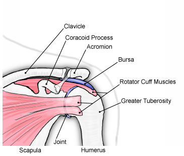
The shoulder is made up of three bones and three joints. What we think of as the shoulder joint is also called the glenohumeral joint, and it is the main ball and socket joint of the shoulder. The ball, or humeral head, is much bigger than the socket, or glenoid. This joint allows the shoulder to move more than any other joint in the body. The socket, or glenoid, is part of the shoulder blade, or scapula. The shoulder blade forms a joint with the rib cage that is also called the scapulothoracic joint. Overall shoulder movement is made up of the movement from both the glenohumeral joint and scapulothoracic joint. The final joint is the connection between the collarbone, or clavicle, and the acromion (part of the shoulder blade) known as the acromioclavicular joint. This joint has a small amount of movement, but is important as the joint acts as a pivot point. A shoulder “separation” involves an injury of this joint.
Capsule
The capsule is the deepest layer of soft tissue and acts as a cover for the shoulder joint. The capsule’s function is to keep the lubrication fluid in the joint and to help support the joint. There are ligaments in the capsule that are thicker parts of the capsule. The ball and socket joint, or glenohumeral joint, and the acromioclavicular joint both have a joint capsule. The joint between the shoulder blade and rib cage, or scapulothoracic joint, does not have a joint capsule.
Ligaments
Ligaments, together with the capsule, act to hold the bones of the shoulder together. There are ligaments between the ball and socket, known as the glenohumeral ligaments, between the collarbone and acromion (part of the shoulder blade), known as the acromioclavicular ligaments, and between the collarbone and the coracoid (part of the shoulder blade), known as the coracoclavicular ligaments. All of these ligaments function to keep the bones in place as the shoulder moves during activity.
Tendons and Muscles
Tendons are made up of elastic and soft connective tissue and they attach muscles to bones. Muscles move the bones by pulling on the tendons. When a muscle is activated or contracts, the tendon pulls on the bone, causing the bone to move. Together, the tendon and muscle form a unit called a muscle-tendon unit. In the shoulder, there is a deep layer of tendons and muscles known as the rotator cuff, and a more superficial layer, made up of the deltoid, pectoralis major, and a number of other muscles of the shoulder, chest, upper back, and neck, that all assist with shoulder motion.
Rotator Cuff
The rotator cuff consists of four muscle-tendon units that originate on the shoulder blade, or scapula, and attach to the tuberosities (bumps of bone) on the ball of the humerus. The role of the rotator cuff is to keep the ball of the humerus centred in the shoulder socket as the shoulder moves through its range of motion and helps to start the movement of the shoulder. The rotator cuff is the primary stabilizer during movement of the ball and socket, or glenohumeral joint. Overuse and traumatic injuries to the rotator cuff are among the most common problems in the shoulder.
Subacromial Bursa
The bursa is a lubricating sac that is between the rotator cuff and the deltoid. It helps the rotator cuff to glide smoothly when the shoulder moves. When the shoulder or rotator cuff is injured or damaged, the bursa can become inflamed, which may lead to pain and a loss of function. This is often referred to as impingement.
Deltoid Muscle
The deltoid is the big muscle that forms a large part of the superficial, or outer layer of the shoulder. It has three parts – the front (anterior), middle, and back (posterior). The deltoid helps to lift the shoulder out sideways (abduction). The front part helps to lift the arm up forwards (flexion) and the back part helps to lift the arm up backwards (extension).
Pectoralis Major
The pectoralis major is a large muscle covering a large portion of the chest, crossing over the shoulder joint, and attaching to the collarbone and humerus. It is part of the superficial, or outer layer of the muscles around the shoulder. The pectoralis major helps with shoulder rotation and flexion, and also helps with posture.
Scapular Stabilizers
The scapular, or shoulder blade, stabilizers consist of muscles around the shoulder blade. The two major muscles are the trapezius and the serratus anterior. Smaller muscles include the rhomboids, the levator scapulae and the latissimus dorsi. The function of these muscles is to stabilize and support the shoulder blade against the rib cage and control its movement. Abnormalities in the movement and rhythm of the shoulder blade are referred to as scapular dyskinesis. These abnormalities may contribute to shoulder pain and are most commonly helped by an active exercise or rehabilitation program that strengthens these muscles.
Visit our Patient Information Resources page for more information and resources.